
Author: Justine Cosman, PT, DPT : Doctor of Physical Therapy, Business Owner, Associate Professor, and Blog Contributor. Explores common client questions and helps find solutions for every day functional health concerns, and then some. Loves empowering others, seeking adventure, and learning every day. Learn more about Justine on Google+.
Are you hurt? You should get that imaged!
I hear that a lot in daily conversation. The expectation that imaging will provide the magic solution that will make us feel better.
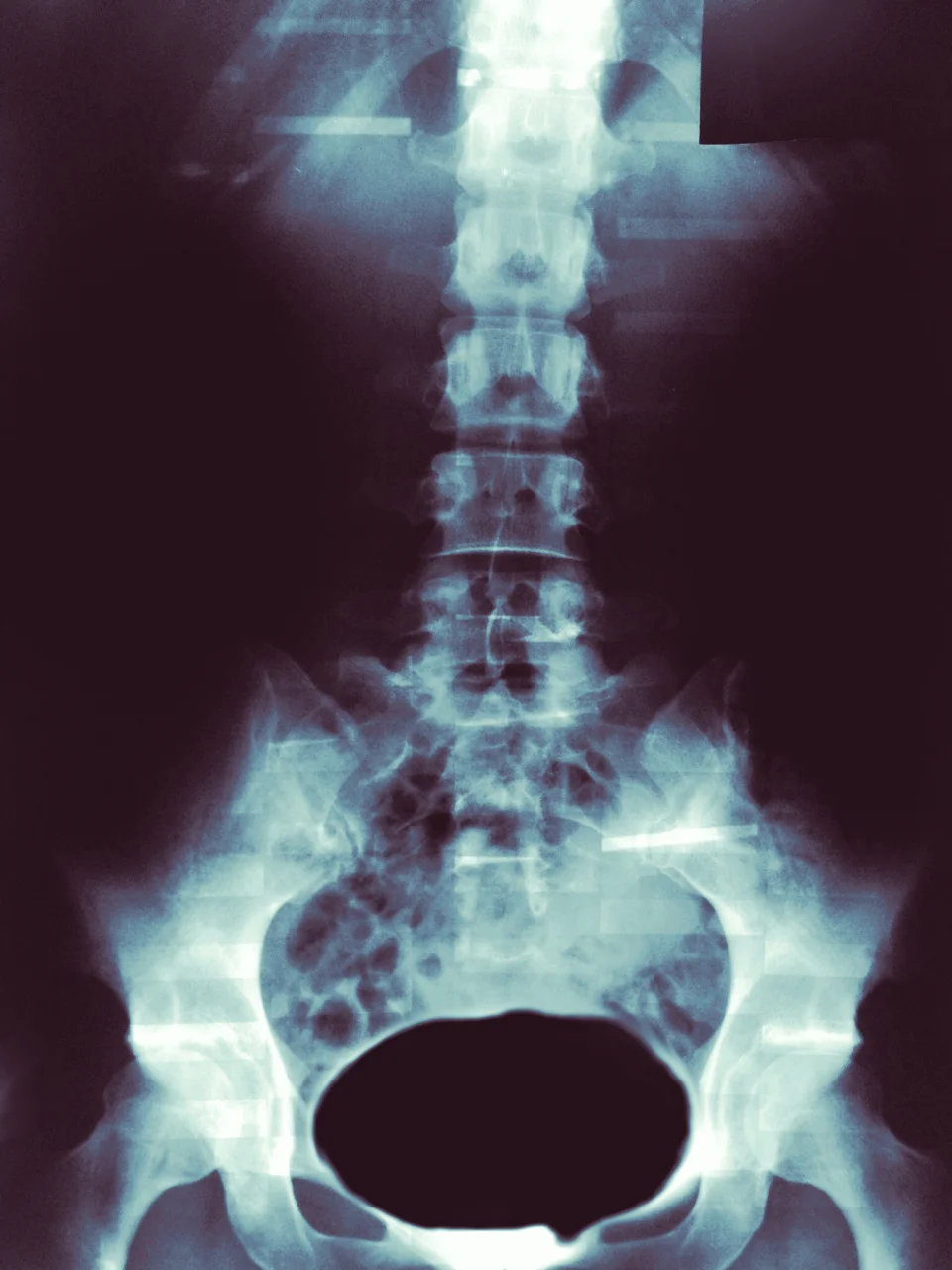
Imaging. It has allowed us to access and see a new world inside of our bodies without cutting beneath the layers. The relative ease of positioning behind an x-ray film or lying down in a tube for an MRI seems very appealing if it allows you to SEE what could be wrong. However, is this really necessary for things such as low back pain or neck pain?
The answer? Actually, no – usually not when they stand alone in assessing the reason for your joint or muscle symptoms.
The reason you should wait? Well, here are a few.
1. These images do not necessarily show you painful structures.
Just because an image shows you that there is a disc herniation or that you have osteoarthritis does not necessarily mean that this is the painful structure. There have been several studies that have shown that when x-rays are performed on non-symptomatic individuals, a large portion of the individuals are found to have typical degenerative processes (such as a “slipped disc” or arthritis), yet they have no pain (1-6, 10-11). This means that for those who are experiencing pain, their image results may be from normal processes and not the true cause of their symptoms. On the other hand, there are other individuals who have significant pain and yet their imaging makes them look as though they are in perfect health.
2. Imaging rarely dictates treatments.
Most often, when a medical professional has screened a client, ruled out other potential causes for symptoms, and suspects a musculoskeletal problem, the next step is conservative treatment. This includes, but is not limited to, physical therapy, massage, acupuncture, and chiropractics. For instance, a study was performed where 2,500 subjects with acute low back pain were imaged. Remarkably, the researchers found that only one x-ray out of the entire group actually changed an individual’s medical treatment. This is less than 1%, yet more than 40% come into the clinic with imaging already performed. (7) Furthermore, recent findings by a collaboration of physician organizations have determined that x-rays should not be performed on patients with low back pain until the pain lasts longer than 6 weeks for this very reason (9).
3. They are an expensive first option.
For around $200 you can get an x ray to peer at your bones and for a little over $5,000 you can get an MRI to get a better look at the soft tissue around those bones (8) – but what do these really tell you if you will likely require conservative treatment first? Physical therapists are skilled at deducing a probable diagnosis based on special test findings during your examination and your personal report of symptoms. With active and passive movement based tests, we are able to work out if it is soft tissue versus skeletal; muscular versus ligamentous; degenerative versus acute.
Are images useful? Of course. They can augment a PT’s exam findings, but they hold little merit when used alone. On the whole, imaging is not necessary for a musculoskeletal injury unless there has been enough trauma to suspect fracture or disc rupture; there are signs that your symptoms may be originating from somewhere outside the musculoskeletal realm (what we term and scan for called “red flags”); if your prior conservative treatment has not helped improve you symptoms over a 4-6 week period (this may vary); and if you are preparing for something big - like surgery.
If you are questioning if imaging is right for you, contact your primary care physician or feel free to contact us. You could save yourself time and money, while getting on the path to better health.
Sources
1. Boden SD et al. “Abnormal magnetic resonance scans of the lumbar spine in asymptomatic subjects: A prospective investigation.” J Bone Joint Surg Am 1990; 72A:403-408
2. Boos N, et al. “1995 Volvo Award in clinical science: The diagnostic accuracy of MRI, work perception, and psychosocial factors in identifying symptomatic disc herniations.” Spine – 1995; 20:2613-2625
3. Boos N, et al. “Natural history of individuals with asymptomatic disc abnormalities in MRI: Predictors of low back pain-related medical consultation and work incapacity.” Spine 2000; 25:1484
4. Borenstein G, Boden SD, Wiesel SW, et al. “The value of magnetic resonance imaging of the lumbar spine to predict low-back pain in asymptomatic individuals: A 7-year follow-up study. J Bone Joint [am] 2001; 83:320-34
5. Jensen MC, et al. “MRI imaging of the lumbar spine in people without back pain.” N Engl J Med – 1994; 331:369-373
6. Powell MC, et al. “Prevalence of lumbar disc degeneration observed by magnetic resonance in symptomless women.” Lancer – 1986; 2:1366-7
7. Unknown. “Investigations for low back and leg pain.” The Physiotherapy Site. http://www.thephysiotherapysite.co.uk/physiotherapy/back-pain/investigations. 08/11/2014.
8. Unknown. "How Much For An MRI? $500? $5,000? A Reporter Struggles To Find Out." Kaiser Health News. http://www.kaiserhealthnews.org/stories/2012/december/09/mri-cost-price-comparison-health-insurance.aspx. 08/11/2014
9. Unknown. Choosing Wisely. http://www.choosingwisely.org. 08/11/2014
10. Weishaupt D et al. “MRI of the lumbar spine: Prevalence of intervertebral disc extrusion and sequestration, nerve root compression and plate abnormalities, and osteoarthritis of the fact joints in Asymptomatic Volunteers.” Radiology – 1998; 209:661-666
11. Wiesel SW, et al. “A study of computer-associated tomography: I. The incidence of positive CAT scans in asymptomatic group of patients.” Spine 1984;9:549-51